STEMI vs Hypertrophy
Reviewed October 2026. Left ventricular hypertrophy (LVH) can produce discordant ST elevation and T-wave changes that mimic acute coronary occlusion. ECG interpretation must take account of QRS voltage, prior tracings, serial changes, symptoms and clinical risk.
ECG example: LVH with secondary repolarisation changes
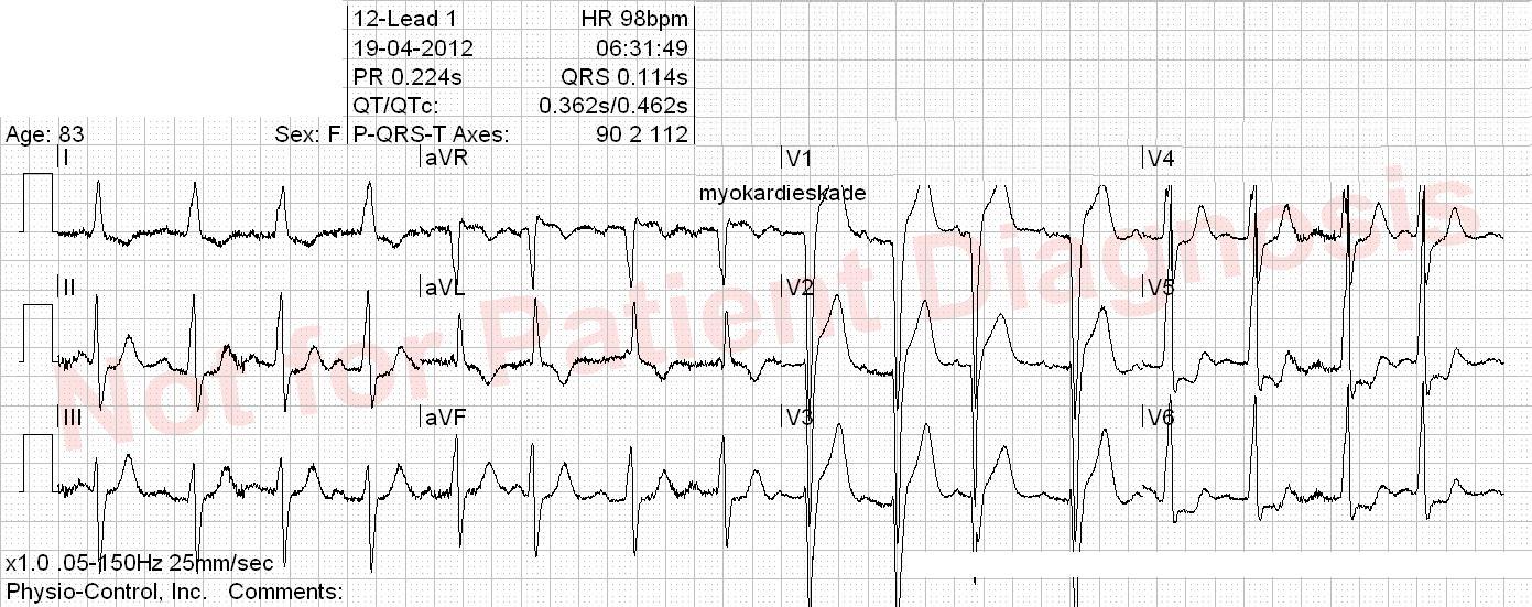
What the Armstrong study found
In a retrospective study of patients with LVH and suspected STEMI, combining ST elevation greater than 25% of QRS peak-to-nadir amplitude with either elevation in three contiguous leads or anterior T-wave inversion improved specificity (reported as 91% versus 58% for conventional thresholds in that study). These performance estimates should not be generalised to all patients or used as a stand-alone rule to withhold urgent angiography. The study requires prospective external validation before adoption as a clinical decision pathway.
Practical interpretation
- Compare the ST segment with expected discordance and examine new dynamic or reciprocal changes.
- Review any previous ECG, evaluate symptoms and haemodynamics, and perform serial ECGs if uncertainty remains.
- Persistent ischaemic pain or a high-risk presentation should trigger urgent specialist assessment even if conventional STEMI criteria are not met.
- Do not apply a fixed 25% LVH ratio as an ESC-endorsed diagnostic or exclusion criterion for acute coronary occlusion.
Original study illustration

Historical study algorithm: illustrative only, not a guideline-validated rule-out tool.
References: Armstrong et al., Electrocardiographic criteria for STEMI in LVH; 2023 ESC ACS guidelines.