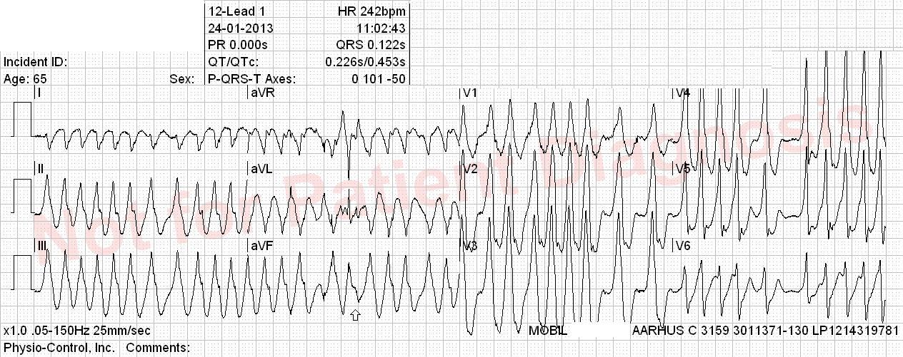
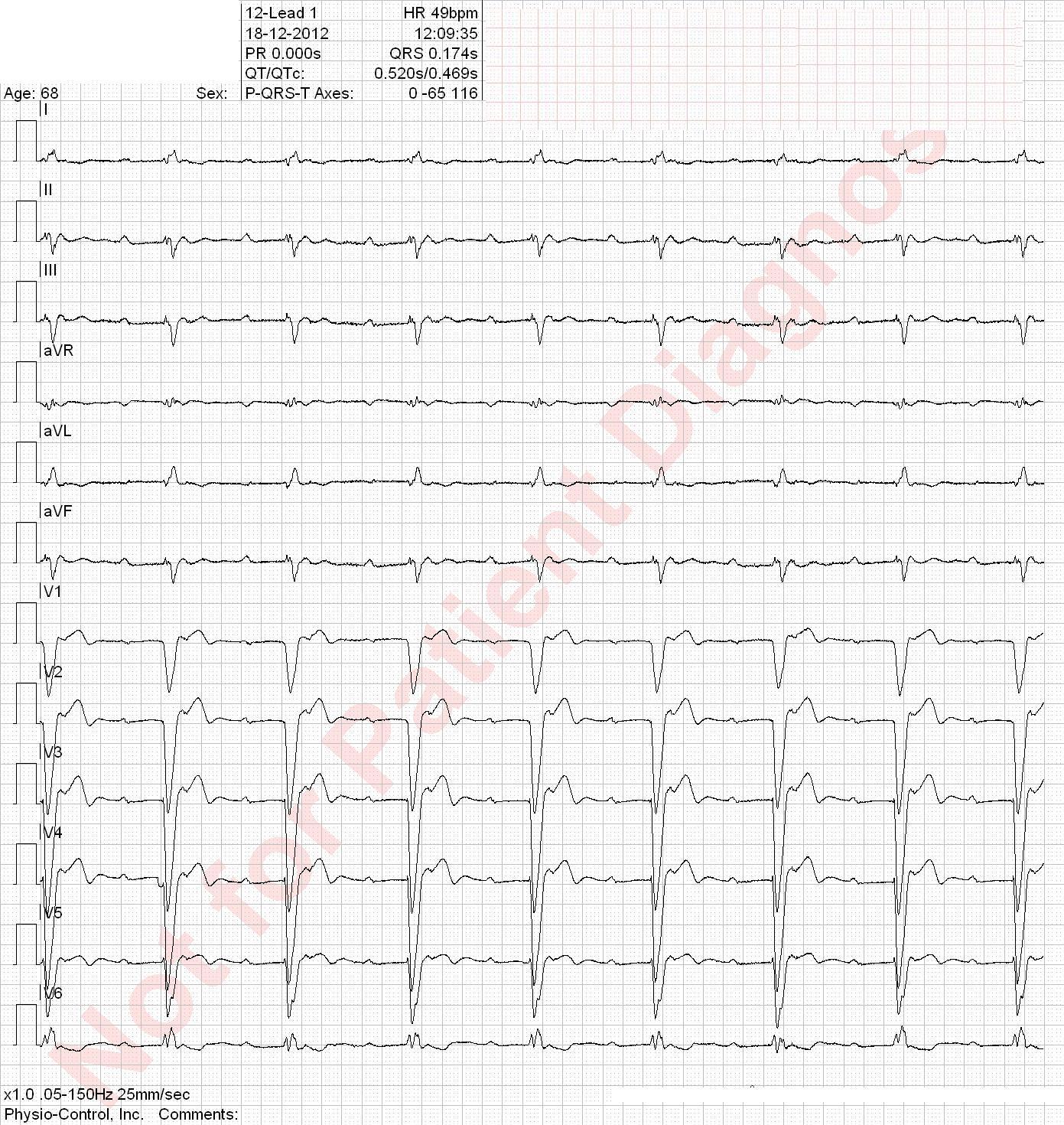
- VT = Ventricular tachycardia
- Afib = Atrial fibrillation
- AVRT = Atrioventricular reentry tachy
- AVNRT= Nodal AVRT
- RBBB = Right bundle branch block
- LBBB = Left bundle branch block
- WPW = Wolff-Parkinson-White syndrome
ECG Interpretation 4
15 March,2015 Antoine AyerQuiz-summary
0 of 51 questions completed
Questions:
- 1
- 2
- 3
- 4
- 5
- 6
- 7
- 8
- 9
- 10
- 11
- 12
- 13
- 14
- 15
- 16
- 17
- 18
- 19
- 20
- 21
- 22
- 23
- 24
- 25
- 26
- 27
- 28
- 29
- 30
- 31
- 32
- 33
- 34
- 35
- 36
- 37
- 38
- 39
- 40
- 41
- 42
- 43
- 44
- 45
- 46
- 47
- 48
- 49
- 50
- 51
Information
This electrocardiogram quiz contains several brief medical histories and their matching ECGs. For each question, you should check between 0 to 4 diagnoses. If the blood pressure is not specified, it means that it is in the normal range.
Remember: Do not use your browser’s “back” button. The result is shown only when you have answered every question.
Enjoy!
You have already completed the quiz before. Hence you can not start it again.
Quiz is loading...
You must sign in or sign up to start the quiz.
You have to finish following quiz, to start this quiz:
Results
0 of 51 questions answered correctly
Your time:
Time has elapsed
You have reached 0 of 0 points, (0)
| Average score |
|
| Your score |
|
Categories
- Not categorized 0%
-
You should improve your ECG skills to optimize your decisions regarding patient triage. You can find interesting educative resources in our link section.
You are welcome to retake the quiz ECG Interpretation 4 or try another EKG Test.
-
Congratulations! You passed “ECG Interpretation 4”.
| Pos. | Name | Entered on | Points | Result |
|---|---|---|---|---|
| Table is loading | ||||
| No data available | ||||
- 1
- 2
- 3
- 4
- 5
- 6
- 7
- 8
- 9
- 10
- 11
- 12
- 13
- 14
- 15
- 16
- 17
- 18
- 19
- 20
- 21
- 22
- 23
- 24
- 25
- 26
- 27
- 28
- 29
- 30
- 31
- 32
- 33
- 34
- 35
- 36
- 37
- 38
- 39
- 40
- 41
- 42
- 43
- 44
- 45
- 46
- 47
- 48
- 49
- 50
- 51
- Answered
- Review
-
Question 1 of 51
1. Question
History of kidney transplantation, known with ischemic heart disease. Unstable angina pectoris for 3 days.
Correct
Hardly meets the criteria for left anterior hemiblock. Axis deviation between -45 ° and -60 °, qR complexes in I and aVL, rS complexes in II, III and aVF, time from onset of QRS to the top of R wave > 45ms (Intrinsicoid deflection).
Incorrect
Hardly meets the criteria for left anterior hemiblock. Axis deviation between -45 ° and -60 °, qR complexes in I and aVL, rS complexes in II, III and aVF, time from onset of QRS to the top of R wave > 45ms (Intrinsicoid deflection).
-
Question 2 of 51
2. Question
This patient has been examined by a practicing cardiologist for suspected tachycardia. He started taking metoprolol. Palpitation for two days.
Correct
Incorrect
-
Question 3 of 51
3. Question
Patient with diabetes and atrial fibrillation. He fell to the floor, was unconscious and had briefly mild twitches. He is now awake but is a little sweaty by the time the ambulance arrived. ABC stable.
Correct
Left anterior fascicular block. Supraventricular bigeminism (atrial tachycardia made of two focus)
Incorrect
Left anterior fascicular block. Supraventricular bigeminism (atrial tachycardia made of two focus)
-
Question 4 of 51
4. Question
Patient with heart failure and COPD. Slowly worsening breathlessness. Cold sweats. No definite chest pain.
Correct
Incorrect
-
Question 5 of 51
5. Question
Correct
Incorrect
-
Question 6 of 51
6. Question
Oppressive left lateral chest pain for 5 hours and impression sleeping left arm. BP 177/95 mmHg. Nitroglycerin spray has some effect on the pain.
Correct
Incorrect
-
Question 7 of 51
7. Question
Correct
The difference between atrial fibrillation (A-fib) and atrial flutter (A-flutter), is clinically relevant because typical flutter can easily be treated by radiofrequency ablation. A-fib and atypical A-flutter requires more expertise and radiofrequency ablation has lower success rate.
Atrial flutter:
Atrial rate ca. 300 bpm (200-400 bpm) with a heart rate typically ca. 150 bpm.
- Typical (type I) atrial flutter: saw-tooth-like waves
- Counterclockwise: negative flutter waves in II, III, aVF and positive in V1
- Clockwise: positive flutter waves in II, III, aVF and negative in V1
- Atypical (type II) atrial flutter: doesn’t fulfill the criteria for typical atrial flutter
Atrial fibrillation:
- No P waves
- Atrial rate typically ca. 400 to 600 bpm
- Heart rate ” Irregularly irregular” and variable (slow A-fib < 60 bpm, fast A-fib > 100 bpm)
Distinguish atypical A-flutter from coarse A-fib:
It may be difficult to distinguish atypical A-flutter from coarse A-fib. ‘Coarse A-fib’ has an “f” wave amplitude> 0.5 mm, which can mimic A-flutter “f” waves morphology. Atypical A-flutter may have varying AV conduction, which can cause an irregular heart rhythm.
BUT:
- A-fib has less organized activity in the atria and thus provides less uniform “f” waves.
- A-fib has an higher atrial rate (400-600 bpm vs 200-400 bpm for A-flutter)
Incorrect
The difference between atrial fibrillation (A-fib) and atrial flutter (A-flutter), is clinically relevant because typical flutter can easily be treated by radiofrequency ablation. A-fib and atypical A-flutter requires more expertise and radiofrequency ablation has lower success rate.
Atrial flutter:
Atrial rate ca. 300 bpm (200-400 bpm) with a heart rate typically ca. 150 bpm.
- Typical (type I) atrial flutter: saw-tooth-like waves
- Counterclockwise: negative flutter waves in II, III, aVF and positive in V1
- Clockwise: positive flutter waves in II, III, aVF and negative in V1
- Atypical (type II) atrial flutter: doesn’t fulfill the criteria for typical atrial flutter
Atrial fibrillation:
- No P waves
- Atrial rate typically ca. 400 to 600 bpm
- Heart rate ” Irregularly irregular” and variable (slow A-fib < 60 bpm, fast A-fib > 100 bpm)
Distinguish atypical A-flutter from coarse A-fib:
It may be difficult to distinguish atypical A-flutter from coarse A-fib. ‘Coarse A-fib’ has an “f” wave amplitude> 0.5 mm, which can mimic A-flutter “f” waves morphology. Atypical A-flutter may have varying AV conduction, which can cause an irregular heart rhythm.
BUT:
- A-fib has less organized activity in the atria and thus provides less uniform “f” waves.
- A-fib has an higher atrial rate (400-600 bpm vs 200-400 bpm for A-flutter)
- Typical (type I) atrial flutter: saw-tooth-like waves
-
Question 8 of 51
8. Question
Patient with COPD and paroxystic atrial fibrillation and has a DDD pacemaker because of sick sinus syndrome. Increasing shortness of breath over a few days. No chest pain.
Correct
Incorrect
-
Question 9 of 51
9. Question
Family history of severe ischemic cardiovascular illness. Active smoking. Onset of chest pain 20 minutes ago.
Correct
Incorrect
-
Question 10 of 51
10. Question
Correct
Incorrect
-
Question 11 of 51
11. Question
Previously healthy patient. Two days with on / off discomfort in the left arm. Tonight at. 0:30 acute onset of severe, retrosternal, oppressive pain radiating to the left arm.
Correct
Incorrect
-
Question 12 of 51
12. Question
Correct
Incorrect
-
Question 13 of 51
13. Question
Previously healthy. 1 ½ hours with strong, oppressive retorsternal chest pain and dyspnea. Good effect of nitroglycerin given prehospitaly. Pale and clammy skin. Has usually a normal ECG.
Correct
Incorrect
-
Question 14 of 51
14. Question
Increasingly poor general condition with weight loss, loathing of food and today also abdominal pain.
Correct
Incorrect
-
Question 15 of 51
15. Question
Correct
Incorrect
-
Question 16 of 51
16. Question
11 days with central chest oppression radiating to both arms as well as sensation of fatigue in both arms.
Correct
Incorrect
-
Question 17 of 51
17. Question
Correct
Incorrect
-
Question 18 of 51
18. Question
Correct
Incorrect
-
Question 19 of 51
19. Question
Correct
Incorrect
-
Question 20 of 51
20. Question
Correct
Incorrect
-
Question 21 of 51
21. Question
Correct
Incorrect
-
Question 22 of 51
22. Question
Correct
Incorrect
-
Question 23 of 51
23. Question
Correct
Incorrect
-
Question 24 of 51
24. Question
Correct
Incorrect
-
Question 25 of 51
25. Question
Cardiac arrest preceded by retrosternal oppression. Single DC shock delivered during transport. Awake.
Correct
Incorrect
-
Question 26 of 51
26. Question
Correct
Incorrect
-
Question 27 of 51
27. Question
Correct
Incorrect
-
Question 28 of 51
28. Question
Former smoker patient with a family history of cardiovascular disease. Over the last few days, he had intermittent pricking sensation in the chest. Marked deterioration one hour ago with the pain radiating now to the neck.
Correct
Incorrect
-
Question 29 of 51
29. Question
Correct
Incorrect
-
Question 30 of 51
30. Question
Dizziness and several episodes of emesis over the past 12 hours. Then retrosternal pain radiating to the neck.
Correct
Incorrect
-
Question 31 of 51
31. Question
Former smoker with hypertension. He woke up this morning with a tingling sensation in both arms. No chest pain. BP 210/93 mmHg. Symptoms disappear completely after nitro spray.
Correct
Incorrect
-
Question 32 of 51
32. Question
Correct
Incorrect
-
Question 33 of 51
33. Question
Patient with COPD and atrial fibrillation treated with digitalis. He complaints of shortness of breath and cough.
Correct
Incorrect
-
Question 34 of 51
34. Question
Correct
Incorrect
-
Question 35 of 51
35. Question
Correct
Incorrect
-
Question 36 of 51
36. Question
Previously healthy woman with a presyncope. Had a “unquiet heart” over the last 50 years but has never been properly investigated.
Correct
Incorrect
-
Question 37 of 51
37. Question
Correct
Incorrect
-
Question 38 of 51
38. Question
Correct
Incorrect
-
Question 39 of 51
39. Question
Previously healthy man without predisposition for heart disease. Brief and mild chest pain 12 hours ago. Have slept and is now pain free.
Correct
Incorrect
-
Question 40 of 51
40. Question
Correct
Incorrect
-
Question 41 of 51
41. Question
Correct
Incorrect
-
Question 42 of 51
42. Question
Correct
Incorrect
-
Question 43 of 51
43. Question
Correct
Incorrect
-
Question 44 of 51
44. Question
Correct
Incorrect
-
Question 45 of 51
45. Question
Correct
Ventricular tachycardia with capture and fusion as well as VA dissociation
Incorrect
Ventricular tachycardia with capture and fusion as well as VA dissociation
-
Question 46 of 51
46. Question
Correct
Incorrect
-
Question 47 of 51
47. Question
Correct
Incorrect
-
Question 48 of 51
48. Question
History of AMI / PCI. Wakes up with oppressive chest pain and feeling of reduced strength in both arms.
Correct
Incorrect
-
Question 49 of 51
49. Question
Correct
Incorrect
-
Question 50 of 51
50. Question
Correct
Incorrect
-
Question 51 of 51
51. Question
Correct
Incorrect


